Your shopping cart is empty!
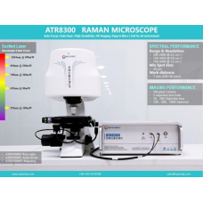
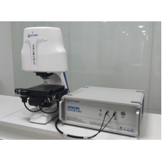
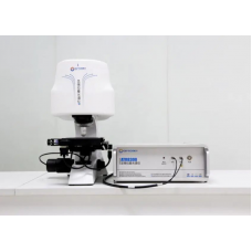
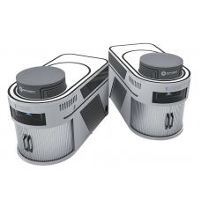
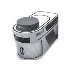
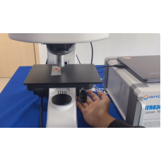
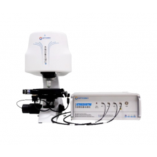
AFM-Raman Microscope


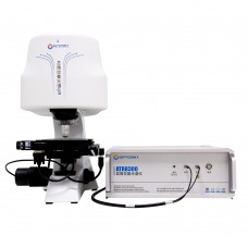
Q: What is the ATRA8300 Raman instrument?
Availability: In Stock
AFM-Raman Microscope
Product Code: ATRA8300
application
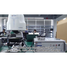
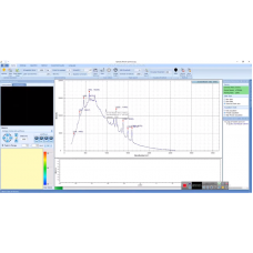
- This Raman instrument integrates an atomic force microscope (AFM), an optical microscope and a laser Raman spectrometer. AFM and Raman spectroscopy can be used respectively to characterize and analyze the surface morphology, particle size, roughness and Raman spectral performance of nanomaterials, thereby providing more comprehensive information on the sample and providing sharp microscopic images. Such integration allows users to improve work efficiency and spend more time on data collection and analysis, truly realizing in-situ detection and analysis of samples. The visual and precise positioning of the Raman detection platform allows observers to detect Raman signals of different surface states on the sample, and can simultaneously display the microdomain shape of the detected location on the computer.
- Microscope objective is specially designed for the Raman system, which makes the laser spot close to the diffraction limit. It overcomes the problem that the focal plane for collecting Raman signals in ordinary Raman systems is slightly higher or slightly lower than the actual optimal focal plane, thus improving the quality of Raman spectra.
- ATRA8300 has no optical path switching moving parts. All optical components are solid-state assembled and work very stably. It perfectly solves the loss of optical path for camera imaging and realizes the separation of camera imaging and Raman signal collection, thereby obtaining the best signal strength.
| Confocal Raman Microscope | |
| Wavelength | 200nm~1100 nm |
| SNR | >6000:1 |
| Dynamic Range | 13000:1 |
| Spectral Range | 250~2700 @ 3-8 cm-1 200~3500 @ 5-10 cm-1 200~4300 @ 6-12 cm-1 Other wavelength ranges can be customized, down to 50 cm-1 |
| Detector | Semiconductor cooling 2048*64 pixel back-illuminated infrared enhanced CCD |
| Pixel | 14 μm * 14 μm |
| Spectral Stability | σ/μ < 0.5% (COT 8 hours) |
| Temperature Stability | Spectral shift ≤ 1 cm -1 (10~40 ℃) |
| Spot Diameter | >1μm |
| Interface | USB 2.0 |
| Microscopic Illumination | LED Kohler illumination |
| Objective Lens | 5X/10X/20X/50X plan apochromatic objective lens |
| Microscope Camera | 5 megapixel CMOS sensor |
| Focus Method | BS: Coarse and fine manual focusing AF, MP: auto focus |
| Laser Stability | σ/μ <±0.2% |
| Laser Linewidth | 0.08 nm |
| Laser Power | >550mW (software adjustable) |
| Laser Wavelength | 785nm (±0.5nm) |
| Operating Mode | Contact mode, tap mode |
| Optional Mode | Friction/lateral force, amplitude/phase, magnetic/electrostatic force |
| Sample Size | Φ≤68mm,H≤20mm |
| Scan Resolution | Horizontal 0.2nm, vertical 0.05nm |
| Scan Rate | 0.6Hz~30Hz |
| Scan Angle | 0~360° |