Your shopping cart is empty!
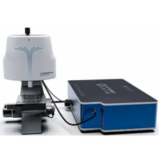
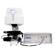
Confocal Raman Microscope for Graphene Analysis


1. What is the ATR8800C Confocal Raman Microscope, and how is it used for graphene analysis?
2. What are the key features of the ATR8800C Confocal Raman Microscope for graphene analysis?
3. How does the ATR8800C help in determining the number of graphene layers?
4. Can the ATR8800C detect defects in graphene?
5. How does the ATR8800C measure stress or strain in graphene?
6. Can the ATR8800C analyze doped graphene?
7. Is the ATR8800C suitable for large-area graphene analysis?
8. What are the advantages of using the ATR8800C for graphene research compared to other Raman microscopes?


Availability: In Stock
Confocal Raman Microscope for Graphene Analysis
Product Code: ATR8800C
application
Optosky ATR8800C Series Confocal Raman Imaging Microscope: Advanced Tool for Comprehensive Graphene Analysis
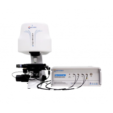
The Optosky ATR8800C Series Confocal Raman Imaging Microscope is a state-of-the-art, high-resolution Raman imaging system specifically designed for graphene research. Whether you're analyzing graphene layers, studying defects, exploring doping effects, or examining stress-induced variations, the ATR8800C provides the precision, speed, and sensitivity required for cutting-edge graphene analysis. By integrating up to four excitation lasers and advanced confocal Raman detection, this microscope enables a comprehensive investigation of graphene’s crystal structure, electronic properties, and mechanical behaviors.
The Optosky ATR8800C Series Confocal Raman Imaging Microscope is a state-of-the-art, high-resolution Raman imaging system specifically designed for graphene research. Whether you're analyzing graphene layers, studying defects, exploring doping effects, or examining stress-induced variations, the ATR8800C provides the precision, speed, and sensitivity required for cutting-edge graphene analysis. By integrating up to four excitation lasers and advanced confocal Raman detection, this microscope enables a comprehensive investigation of graphene’s crystal structure, electronic properties, and mechanical behaviors.
Precision in Graphene Layer Characterization
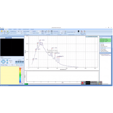
The ATR8800C Confocal Raman Imaging Microscope excels in the precise layer-by-layer analysis of graphene. By utilizing the distinct G peak (around 1580 cm⁻¹) and 2D peak (around 2700 cm⁻¹) of graphene's Raman spectrum, the ATR8800C provides unparalleled accuracy in determining the number of layers present. For monolayer graphene, the 2D peak appears as a sharp, symmetric feature, while for bilayer or multilayer graphene, the peak broadens and becomes asymmetric.
The ATR8800C’s high spatial resolution (<1 μm) allows researchers to measure these Raman features with precision, differentiating between monolayer, bilayer, and multilayer graphene. By analyzing the 2D peak position and Full Width at Half Maximum (FWHM), the ATR8800C provides exact measurements of the layer thickness and enables researchers to map out the layer distribution across the entire sample with ease.
Defect Analysis and Mapping in Graphene
The D peak (around 1350 cm⁻¹) and G peak (around 1580 cm⁻¹) are critical for analyzing defects in graphene. The intensity ratio of the D peak to the G peak (ID/IG) provides valuable insight into the defect density of the material. Using the ATR8800C, researchers can perform high-resolution defect mapping to visualize the spatial distribution of defects, such as vacancies, doping-induced defects, or grain boundaries, across the graphene sample.
The ATR8800C’s advanced capabilities allow for the detection of even small-scale defects, with the system providing precise spatial mapping of the ID/IG ratio. This enables detailed studies of the material’s quality and structural integrity, which is crucial for optimizing graphene for various applications in nanoelectronics, sensors, and energy storage devices.
Doping Effects and Electronic Structure Analysis
Doping plays a significant role in altering graphene’s electronic properties, and the ATR8800C Confocal Raman Imaging Microscope is an ideal tool for studying these effects. Whether you're investigating n-type or p-type doping, the ATR8800C provides non-destructive, high-resolution measurement of doping-induced shifts in the G peak and 2D peak.
Through precise analysis of the G peak shift (blue shift for n-type and red shift for p-type doping), as well as changes to the 2D peak, the ATR8800C offers insights into the Fermi level shifts and charge carrier density in graphene. These measurements are critical for understanding how doping affects carrier mobility and electronic transport properties, enabling the optimization of graphene for high-performance electronic devices, transistors, and sensor technologies.
Stress and Strain Analysis in Graphene
Graphene’s mechanical properties can be significantly influenced by applied stress. The ATR8800C Confocal Raman Imaging Microscope allows for precise stress analysis by detecting shifts in the G peak and 2D peak positions that result from tensile or compressive stress. Even slight mechanical deformation can induce significant Raman spectral changes, making it possible to monitor strain in graphene at the micro-scale.
With its high spatial resolution, the ATR8800C enables stress mapping across a graphene sample, providing detailed insights into strain distribution and its relationship to the material's electronic properties. This capability is invaluable for applications in nanoelectronics, where stress management is crucial for device performance and reliability.
Substrate Effects on Graphene’s Raman Spectrum
The interaction between graphene and its underlying substrate can significantly alter its Raman spectrum. The ATR8800C Confocal Raman Imaging Microscope is equipped to study these substrate-induced effects, whether the graphene is deposited on SiO₂/Si, SiC, or other materials.
Using Raman spectroscopy, the ATR8800C can measure shifts in the G peak and 2D peak positions, revealing how the substrate influences graphene’s electronic structure, layer stacking, and doping levels. This information is essential for optimizing graphene-based devices in fields such as optoelectronics and photodetectors, where the choice of substrate plays a critical role in device performance.
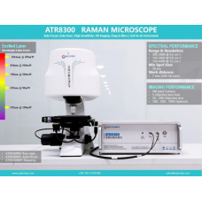
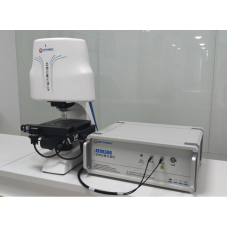
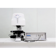
Unmatched Sensitivity, Resolution, and Automation
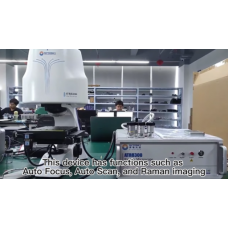
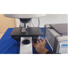
The ATR8800C Series offers superior Raman sensitivity with a signal-to-noise ratio (SNR) > 6000:1, enabling the detection of even the faintest Raman signals with unmatched clarity. The system’s automatic focusing and auto-scanning capabilities allow for seamless, high-speed imaging with one-click operation, making it ideal for high-throughput analysis of graphene samples.
With a 100×100 mm imaging range and the ability to perform automated imaging splicing, the ATR8800C can rapidly map large areas of graphene samples while maintaining high spatial resolution. The inclusion of high-quality objective lenses and a 5 million-pixel camera ensures that every detail is captured with precision and clarity, providing comprehensive Raman data in real-time.
Conclusion: The Ideal Tool for Graphene Research
The Optosky ATR8800C Confocal Raman Imaging Microscope is an indispensable tool for graphene research and advanced material science. With its superior sensitivity, high spatial resolution, and versatile wavelength options, the ATR8800C empowers researchers to conduct detailed structural, electronic, and mechanical analyses of graphene. Whether you're exploring graphene layers, studying defects, analyzing doping effects, or examining stress-induced changes, the ATR8800C provides the accuracy, speed, and reliability needed for cutting-edge research.
The Optosky ATR8800C Confocal Raman Imaging Microscope is an indispensable tool for graphene research and advanced material science. With its superior sensitivity, high spatial resolution, and versatile wavelength options, the ATR8800C empowers researchers to conduct detailed structural, electronic, and mechanical analyses of graphene. Whether you're exploring graphene layers, studying defects, analyzing doping effects, or examining stress-induced changes, the ATR8800C provides the accuracy, speed, and reliability needed for cutting-edge research.
| Confocal Raman Microscope | |
| Excitation Wavelength | 266、325、532、638、785、 1064nm Optional, integrate up to 4 excitation wavelengths simultaneously |
| Detector | 1) Deep cooling area array CCD: 2000X256 pixels 2) Deep cooling high sensitivity EMCCD: 1600X200 pixels 3) Deep cooling area array InGaAs CCD: 512X1 pixels Up to 2 detectors can be integrated, choose one from detector 1# or detector 2#; |
| Spot Diameter | >1μm |
| Working Humidity | constant humidity (50±10%) |
| Working Temperature | Constant temperature (25±2℃) |
| Interface | USB3.0 |
| Microscopic Illumination | High brightness and long life white LED |
| Microscope Camera | 5 million pixel industrial camera |
| Focus Method | conjugate focus |
| Laser Stability | σ/μ <±0.2% |
| Laser Power | 266nm:30 mW 325nm:30mW 532nm: 100mW 633nm:80mW 638nm:80mW 785nm:350mW 1064nm:500mW |
| Voltage | 100 ~240 VAC |
| Peak Power | < 200 W |
| Dimensions | ATR8800-FL350:905(L)×58.3(W)×643(H) ATR8800-FL510: 1009(L)×58.3(W)×643(H) ATR8800-FL810: 1520(L)×68.3(W)×643(H) |
| Weights | ATR8800-FL350:59 Kg ATR8800-FL510:63 Kg ATR8800-FL810:78 Kg |
Multi-Wavelength Flexibility:
Supports up to 4 excitation wavelengths: 266 nm, 325 nm, 514 nm, 532 nm, 638 nm, 785 nm, 830 nm, and 1064 nm, enabling versatile material analysis with a choice of single, dual, triple, or quad-band configurations.
Superior Spatial Resolution:
Achieves <1 μm laser spot size, providing high spatial resolution for precise imaging of microstructures, ideal for graphene, nanomaterials, and thin films.
Ultra-High Spectral Resolution:
Offers an ultra-high spectral resolution of 0.35 cm⁻¹, enabling detailed chemical analysis and accurate detection of material properties at the molecular level.
Automatic Focus and Scan:
Features fully automatic focusing and auto-scanning, allowing for quick, hands-free operation with high-speed, high-precision imaging of micron-sized specimens in under 2 seconds.
Advanced Optical Design:
Equipped with Raman-specific objective lenses that ensure diffraction-limited laser spots, enhancing the signal-to-noise ratio and improving spectral stability.
High Sensitivity:
Delivers SNR > 6000:1, enabling the detection of faint Raman signals with exceptional sensitivity for low-concentration or trace material analysis.
High-Resolution Camera:
5 million-pixel camera ensures real-time visualization with crystal-clear images, providing accurate monitoring of Raman signals during analysis.
Large Imaging Range:
Supports automated imaging splicing for large-area scans of up to 100×100 mm, providing comprehensive Raman data over broad sample areas.
Exclusive Sealed Sample Compartment:
Closed hatch design allows for continuous, light-efficient measurements without turning off lab lights, ensuring a stable environment for accurate analysis.
Enhanced Software Integration:
The intuitive software offers multi-band spectral splicing, advanced data analysis, and large-area imaging capabilities, enabling users to analyze complex datasets seamlessly.
Non-Destructive and Fast:
A powerful non-destructive technique for quickly measuring intrinsic properties of materials, such as graphene layers, defects, doping, and stress.
Rotated Grating for Spectral Precision:
Utilizes a rotated grating for enhanced spectral range and accuracy, ensuring precise wavelength selection for high-quality Raman spectra.
- Nanoparticles and new materials
- Research institute research
- Biology
- Forensic Medicine Identification
- Material science
- Medical Immunoassay
- Agriculture and food identification
- Gem and inorganic mineral identification
- Environment
ATR8800C Confocal Raman Microscope for Graphene Analysis
1. What is the ATR8800C Confocal Raman Microscope, and how is it used for graphene analysis?
2. What are the key features of the ATR8800C Confocal Raman Microscope for graphene analysis?
- High Spatial Resolution: The ATR8800C provides high spatial resolution (up to 0.1 µm), enabling precise layer thickness determination and defect mapping of graphene samples.
- Confocal Imaging: With confocal imaging, the microscope isolates specific layers or areas within a sample, allowing detailed mapping of graphene properties.
- Comprehensive Spectral Range: It covers the full range of Raman peaks for graphene analysis, including G, 2D, D, and D' peaks, essential for defect detection and layer number determination.
- Non-Destructive Testing: The ATR8800C uses Raman spectroscopy, which is a non-invasive technique, making it ideal for analyzing fragile graphene without altering or damaging the material.
- Advanced Mapping Capabilities: The microscope can generate detailed Raman maps that visualize defects, strain, and layer variations in graphene at micro and nanoscales.
3. How does the ATR8800C help in determining the number of graphene layers?
4. Can the ATR8800C detect defects in graphene?
5. How does the ATR8800C measure stress or strain in graphene?
6. Can the ATR8800C analyze doped graphene?
7. Is the ATR8800C suitable for large-area graphene analysis?
8. What are the advantages of using the ATR8800C for graphene research compared to other Raman microscopes?
- Higher Resolution: The ATR8800C provides superior spatial resolution (down to 0.1 µm), making it ideal for high-precision measurements of graphene layers and defects.
- Advanced Mapping: Its ability to create Raman maps of large areas with high resolution is unparalleled, enabling detailed analysis of stress, defects, and layer variations across the entire graphene sample.
- Non-Destructive Testing: Unlike other techniques (e.g., electron microscopy), Raman spectroscopy is completely non-destructive, allowing for repeated measurements without damaging the sample.
- Comprehensive Analysis: The ATR8800C provides detailed data on layer thickness, defect density, doping levels, and stress, all in a single system.
The ATR8800C Confocal Raman Microscope is a high-performance, non-destructive analytical instrument that uses Raman spectroscopy to examine the molecular and structural properties of materials, including graphene. The microscope combines high-resolution confocal imaging with advanced Raman spectroscopic capabilities, making it ideal for precise graphene layer analysis, defect detection, doping characterization, and stress analysis.
For graphene, the ATR8800C can accurately identify layer thickness, detect defects (such as vacancies, grain boundaries, and doping), and assess strain or stress in graphene sheets. This is essential for optimizing graphene’s properties for applications in electronics, sensors, and advanced materials.
The ATR8800C Confocal Raman Microscope can precisely determine the number of graphene layers based on the characteristics of the Raman spectra. The key peaks used for layer identification are the G peak (around 1580 cm⁻¹) and the 2D peak (around 2700 cm⁻¹). For monolayer graphene, the 2D peak is sharp and symmetric, whereas for bilayer or multilayer graphene, the 2D peak becomes broader and asymmetric.
The ATR8800C allows researchers to measure the 2D peak's shape, position, and full width at half maximum (FWHM) with high accuracy, making it possible to distinguish between monolayer, bilayer, and multilayer graphene. This is essential for applications where the number of graphene layers affects the material’s electronic properties.
Yes, the ATR8800C Confocal Raman Microscope is an excellent tool for detecting defects in graphene. Defects in graphene, such as vacancies, grain boundaries, and dopants, manifest as changes in the Raman spectrum—specifically in the D peak (around 1350 cm⁻¹) and G peak (around 1580 cm⁻¹).
The D/G peak intensity ratio (ID/IG) is commonly used to assess defect density in graphene. The ATR8800C’s high sensitivity allows for precise detection of even small defects, providing an in-depth view of the quality of graphene. It can also identify different types of defects (e.g., edge defects and sp³ hybridized defects) by measuring other Raman peaks like the D' peak and G' peak.
The ATR8800C Confocal Raman Microscope is capable of measuring stress and strain in graphene by analyzing shifts in the Raman peaks. When graphene is subjected to stress (compressive or tensile), the G peak and 2D peak shift in position. Typically, compressive stress leads to a blue shift (toward higher wavenumbers), while tensile stress causes a red shift (toward lower wavenumbers).
Using Raman mapping, the ATR8800C can visualize how stress is distributed across a graphene sample, providing valuable information about strain engineering and the material's mechanical properties. This is especially important for nanoelectronics and sensors, where the stress-induced properties of graphene can affect device performance.
Yes, the ATR8800C Confocal Raman Imaging Microscope is well-suited for analyzing doped graphene, including both n-type and p-type doping. Doping affects the Fermi level of graphene, leading to shifts in the Raman spectrum, especially in the G peak and 2D peak.
In n-type doping, the G peak exhibits a blue shift, while in p-type doping, the 2D peak shows a red shift. The ATR8800C can detect these shifts with high precision, enabling researchers to quantify dopant levels and understand how doping affects graphene's electronic structure and properties. This makes the ATR8800C ideal for graphene-based sensors, transistors, and other electronic devices.
Yes, the ATR8800C Confocal Raman Microscope is ideal for large-area graphene analysis. With its advanced Raman mapping capabilities, it can scan large areas of a graphene sample (even several millimeters) and create detailed Raman maps that highlight variations in layer number, defect density, and stress distribution across the sample.
The high spatial resolution of the ATR8800C ensures that these large-area scans maintain excellent detail, making it suitable for applications that require extensive analysis, such as graphene production and quality control in commercial graphene manufacturing.
The ATR8800C Confocal Raman Microscope offers several advantages for graphene research:
These advantages make the ATR8800C the top choice for graphene researchers and engineers working in materials science, nanoelectronics, sensors, and other advanced fields.