Your shopping cart is empty!
UV Fluorescence Hyperspectral Imager
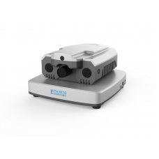
ATHF5010 is an auto-focusing and auto-scanning microscopic fluorescence hyperspectral imager carefully developed by AOPU Tiancheng. Its excitation light is ultraviolet light or blue light. Fluorescence spectroscopic testing of the location. It can both perform spatial imaging and scan out the fluorescence spectrum at each location.
Availability: In Stock
UV Fluorescence Hyperspectral Imager
Product Code: ATHF5010
application
Description
ATHF5010 is an auto-focusing and auto-scanning microscopic fluorescence hyperspectral imager carefully developed by AOPU Tiancheng. Its excitation light is ultraviolet light or blue light. Fluorescence spectroscopic testing of the location. It can both perform spatial imaging and scan out the fluorescence spectrum at each location.
A snapshot can obtain the spatial and fluorescence spectral information of the linear region within 100 ms, and the fluorescence hypercube data of the sample can be obtained by scanning the electric displacement stage. Covering the UV-Vis band with high spectral resolution, it is suitable for mission-critical applications ranging from biomedicine to forensic science to UV measurement.
ATHF5010 is equipped with a 100mm large-area motorized scanning platform, supplemented by an advanced and fast ultra-large image stitching algorithm, so as to achieve the functions of fast scanning and large-area imaging.
Features
- Excitation wavelength: 275, 310, 365 or 405nm, others can be customized
- Auto Focus, Auto Scan
- Optimum number of spatial channels: 2048
- Maximum number of spectral channels: 2048 channels
- Linear push-broom imaging
- Full-band fluorescence and spatial simultaneous imaging
- Optimal fluorescence spectral resolution: better than 1.5nm
- Transmission grating spectrum, higher sensitivity
- 5 million pixel high-definition visible light imaging
- 5-hole microscope stand, motorized switching
- Novel integrated frame provides excellent stability and operability
Parameters | ATHF5010 |
Excitation parameters | |
Excitation wavelength | Two of 275, 310, 365, 405nm, other excitation wavelengths can be customized |
Excitation light source | Long-life UV LED light source, UV laser light source is optional |
Maximum excitation power | 25W, optional 500W |
Fluorescence receiving part parameters | |
Spectral detection range | 400-1000 nm range Full Spectrum |
Maximum Number of Fluorescent Channels | 2048 |
Fluorescence spectral resolution(FWHM) | 1.5 nm |
Number of imaging space channels | 2048 |
Best spatial resolution | 1 μm (In case of 100x objective lens) |
Detector | Cooled detector |
Detector resolution | 2048X2048 |
Refrigeration temperature | 10℃ |
Integration time | 1ms-30min |
Detector interface | USB 3.0 |
Dynamic Range | ≥60dB |
Maximum frame rate | 120Hz |
Visible light imaging system | |
Light source | LED White light source |
Imaging camera | 500 megapixel digital camera |
Camera interface | USB2.0 |
Microscope & Stage | |
Microscope light path | Infinity optical path |
Objective lens mounting plate | 5 holes, electric switching |
Objective lens | 5x、10x、20x、50x ,also 100x optional |
Focusing device | Manual focus or automatic focus, and a focus upper limit device |
Stage | Steel wire drive stage (X axis does not protrude), double clamp structure |
Stage area | 220X200mm |
Scanning electronic control platform | |
Range of movement | 300 mm Scanning itinerary, other itineraries can be customized |
Mobile resolution | 0.1 μm |
Positioning accuracy | 1 μm |
Scanning speed | 20mm/s |
Focus method | Manual, electric, real-time focus |
Z axis (electric control, auto focus) | |
Focus accuracy | ≤ ±0.2 μm |
Maximum stroke | 100 mm |
Focus speed | Does not exceed10 s |
Dimensions | 650 X 520 X 520 mm |
Weight | 49.3 kg |
Software part | |
Function | Visual imaging and real-time fluorescence spectral detection |

-600x600.jpg)
-74x74.jpg)


