Your shopping cart is empty!
Fluorescence Imaging Microscope
Availability: In Stock
Fluorescence Imaging Microscope
Product Code: ATF8000
application
Description
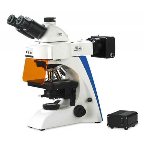
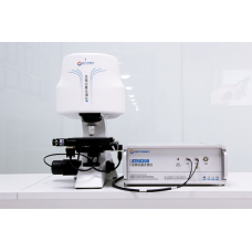
ATF8000 is a large-area fluorescence imaging microscope with auto-focus and auto-scan carefully developed by Optosky. It uses a 200W ultra-high pressure mercury lamp as the light source and uses a 3-stage filter to filter the light source. It can output 3-color images. Excitation light; for emission light, 4-5 sets of filter wheels are used to collect fluorescence signals of different bands. ATF8000 is equipped with a 50X50mm large-area motorized scanning platform, supplemented by an advanced and fast ultra-large image stitching algorithm, so as to achieve the functions of fast scanning and large-area imaging. ATF8000 is equipped with a high-stability autofocus system, which can dynamically adjust the focus of the target in real time to achieve the best imaging effect. ATF8000 is connected to the computer through the USB 2.0 interface, and there is also advanced and easy-to-use PC-side control software, which can achieve perfect experimental operation.
Features
- Powerful image acquisition and analysis software
- Superior infinity chromatic aberration corrected optical system ensures excellent resolution and clarity
- Six-hole turntable fluorescence device, providing a variety of fluorescence excitation block options
- Five-hole turntable phase contrast device, equipped with 10X/20X/100X infinity Plan-field phase-contrast objective for phase-contrast and bright-field observation
- Large area automatic scanning, automatic image stitching, real-time autofocus
- Novel integrated frame provides excellent stability and operability
- Modular structure design, multi-functional combination to ensure the versatility of the system
- Super large field of view eyepiece, the field of view can reach 23mm
- Two-speed conversion trinocular observation tube, 100% observation; 20% observation, at the same time 80% photography
Parameters | Specifications |
Optical system | OTICS Infinity Chromatic Aberration Corrected Optical System |
Magnification range | 4 0X ~ 1600X |
Eyepiece | 10 X large field of view, high eye point plan eyepiece, field of view Φ22 mm (Φ23 mm optional ) |
Infinity Plan Achromats mirror | 2.5X / 4X /10X / 20X ( S ) /40X ( S ) / 60X ( S ) / 100X ( S , O ) / 100X ( S , W ) |
Observation tube | Hinged trinocular observation tube, 30° tilt , interpupillary distance adjustment 48 mm ~ 76 mm , trinocular two-speed conversion |
Converter | internal positioning five-hole converter |
Focusing device | Coarse and fine coaxial focusing, coarse adjustment with tightness adjustment, and a focus upper limit device |
Stage | Steel wire transmission stage ( X- axis does not protrude), double-piece clamp structure |
Condenser | N . A . 0.9/0.13 Swing-out Condenser with Iris Stop |
lighting system | 6 V /30 W halogen lamp (wide voltage input: 100V ~ 240V), field of view diaphragm, center adjustable |
| 1 00 W digital mercury lamp power epifluorescence device, optional B , G , Uv , V and others special filter |
LED fluorescent power supply, optional B , G , Uv , V color filters | |
Phase contrast device | Five-hole turntable phase contrast device (10 X /20 X /40 X /100 X infinity plan phase contrast objective lens) |
Independent phase contrast device | |
Dark field device | Dark field condenser (dry, wet) |
Polarizer | polarizer, polarizer |
Camera device | Equipped with 320/5 million pixel digital camera system for bright field photography |
configuration 3.1/5.1 million pixel CCD digital camera for professional picture recording | |
X , Y axis electric control two-dimensional platform | |
Maximum imaging range | 50 X 50 mm |
Mobile resolution | 0.1μ _ m |
Positioning accuracy | 1 μm _ |
Scanning speed | 20mm / s _ |
Focus method | Motorized , real-time focus |
Z axis (electric control, auto focus) | |
Focus accuracy | ≤ ±0.2μm |
Maximum stroke | 20 mm |
Focus speed | no more than 10 the s |
Dimensions | 2 90 X 210 X 220 mm |
Weight | 1 2 .3 kg |

