Your shopping cart is empty!
Auto-focus,Auto-scan Super FOV Fluorescence Microscopic


Availability: In Stock
Auto-focus,Auto-scan Super FOV Fluorescence Microscopic
Download Data Sheet
Product Code: ATF8100
application
What are the advantages of choosing the ATF8100 Auto-focus, Auto-scan Super FOV Fluorescence Microscopic?
ATF8100 is an Auto-focus, Auto-scan Super FOV Fluorescence Microscopic designed by Optosky,two light sources are available,100W digital mercury lamp power supply and LED fluorescent light source. A third-order filter is used to filter the light source, and a six-hole turntable epi-fluorescence device (optionally with B, G, UV, and V filters) can be used to switch between different color filters to collect fluorescent signals in different wavelength bands.
The ATF8100 is TE-cooled down to -20℃, high sensitivity, and high-resolution spectrometer, which can perform spectral analysis on the target in the imaging area with a resolution of <2nm.
ATF8100 is loaded with a 50X50mm large-area electric scanning platform, supplemented by advanced and fast super-large image stitching algorithms, thus achieve the functions of rapid scanning and large-area imaging.
The ATF8100 is equipped with a highly stable autofocus system that can dynamically adjust the focal length of the target in real-time to achieve the best imaging effect.
The ATF8100 is connected to the computer via a USB 2.0 interface and has advanced and easy-to-use PC-side control software, which can achieve perfect experimental operation.
Model | Explanation |
ATF8100 | 5-mega pixels CCD |
ATF8100A | TE-COOLED 20-mega pixels high performance sCMOS, -15℃, sensitivity increased by 50% |
- Schematic diagram of fluorescence spectroscopic imaging microscope
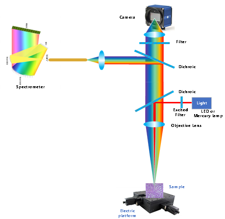
Figure1. Schematic diagram of fluorescence spectroscopic imaging microscope
- Epi-fluorescence intermediate and spectrum collection intermediate
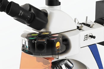
Figure2. Epi-fluorescence intermediate (left) and spectrum collection intermediate (right)
| Detector | |
| Detector type | 2048 pixels CMOS |
| Sensitivity | 1300 V/(lx·s) |
| Dark noise | 0.4mV/RMS |
| Optical parameter | |
| Signal-to-noise | >800:1 |
| Optical Design | f/4 cross asymmetric C-T optical path |
| Raman Instrument | |
| Integration time | 1ms-60min |
| Spectral Range | 300-1100nm, 200-400nm, 500-1100nm, 350-810nm Customzed |
| Resolution | 1-2.5nm |
| Dynamic Range | 10000:1 |
| Focusing | Electric, real-time focusing |
| Raman Spectrometer System Parameters | |
| Interface | SMA905 |
| Epi-fluorescence system | |
| Light source | 100W digital mercury lamp power supply or LED fluorescent light source (choose one) |
| Six-hole tumtable epi-fluorescence device | Standard three-channel switching: blue excitation B, green excitation G, purple |
| Excitation filter set | Blue excitation wavelength:450~490nm Emission wavelength:515nm Green excitation wavelength:495~555nm Emission wavelength:595nm Violet excitation wavelength:380~415nm Emission wavelength:475nm (three channels) |
| Microscopic optical system | |
| Optical system | OTICS infinite distance chromatic aberration correction optical system |
| Magnification range | 40X~1600X |
| Eyepiece | 10X wide field of view, high eyespots flat field eyepiece,field of view Φ22mm (Φ23mm optional) |
| Infinite distant flat field achromatic objective lens | Standard configuration 4X/10X/20X/40X (other optional) |
| Observation tube | Hinged trinocular observation tube, tilted at 30°, interpupillary distance adjusted from 48mm to 76mm, three eyepieces and two gear shifts |
| Converter | Internal tilt type internal positioning five-hole converter |
| Focusing device | Coarse and micro coaxial focus adjustment, coarse adjustment belt elastic adjustment, and the focus of the upper limit device |
| Micrroscope stage | Steel wire transmission stage (X axis does not protrude), double clip structure |
| Focusing mirror | N.A.0.9/0.13 Swing-out focusing mirror,with variable light bar |
| Transmission lighting system | 6V/30W Halogen lamp(Wide voltage input:100V~240V),Field light bar, adjustable center |
| Camera | Equipped with 320/5 megapixels and other digital camera system for bright field shooting Equipped with 310/5.1 megapixel CCD digital camera for professional picture shooting |
| X,Y-axis electronic controlled platform | |
| Moving Range | 50 X 50 mm |
| Moving Resolution | 0.1 μm |
| Positioning Accuracy | 1 μm |
| Scan Speed | 20mm/s |
| Z-axis(Auto-Focus) | |
| Focus Accuracy | ≤ ±0.2 μm |
| Max distance | 20 mm |
| Focus Speed | No more than 10 s |
| Dimensions | 290 X 210 X 220 mm |
What are the major features of the ATF8100 Auto-focus, Auto-scan Super FOV Fluorescence Microscopic?
l TE-cooled down to -20℃, high sensitivity and a high-resolution spectrometer
l Large area automatic scanning, automatic image splicing
l Real-time autofocus
l Powerful image acquisition and analysis software
l Excellent infinity chromatic aberration correction optical system to ensure excellent resolution and clarity
l Six-hole rotating disk fluorescence device provides a variety of fluorescent excitation block selection
l Five-hole turntable phase-contrast device, equipped with 4X/10X/20X/40X/100X and other infinite flat field phase contrast objectives, can be used for phase contrast and bright field observation
l Novel integrated frame provides excellent stability and operability
l Modular structure design, multi-functional combination to ensure the versatility of the system
l Large visual field eyepiece, the field of view up to 23mm, more flat and comfortable observation
l Two - shift three - eye observation tube,100% observation; 20% observation, 80% photography at the same time
What are the applications of the ATF8100 Auto-focus, Auto-scan Super FOV Fluorescence Microscopic?
l Research Lab
l Hospital and Biochemical Lab
l Hospital Clinical Test
l University Teaching